We can all agree dog breath leaves little to be desired, but it does keep our furry friends alive – so I guess we can live with it.

The canine respiratory system, like ours, is responsible for taking in oxygen and subsequently releasing wastes like carbon dioxide. Unlike us however, dogs do not sweat through the skin, which means the respiratory system also plays a crucial role in temperature regulation. The lungs are also highly susceptible to airborne viruses and irritants, which is why it’s so important to understand how your dog’s respiratory system works.
Below is an overview of the canine respiratory system. Take a deep breath, because we’re divin’ in!
The Anatomy of the Canine Respiratory System (An Overview)
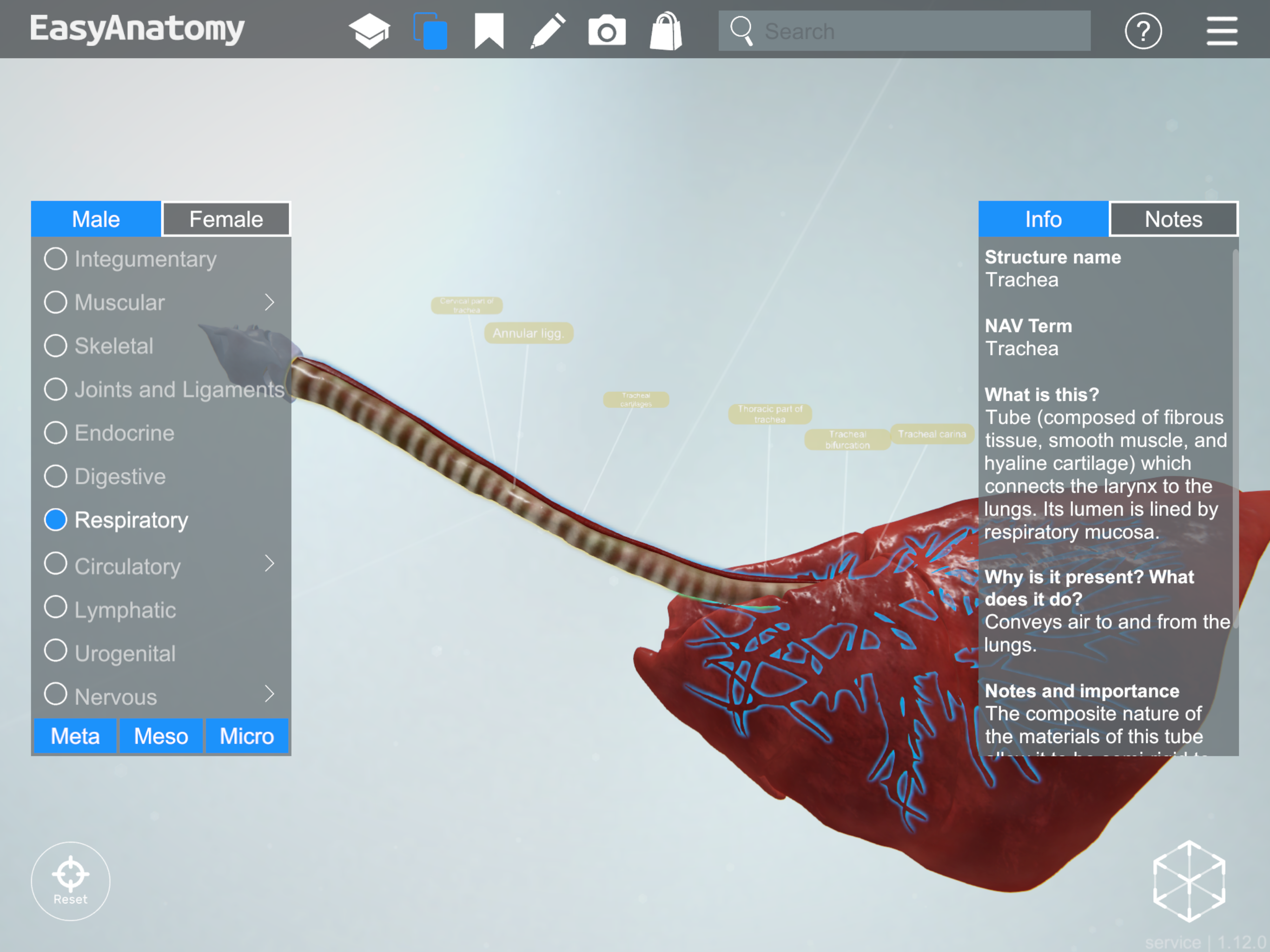
Trachea
NAV Term: Trachea
What is this? Tube (composed of fibrous tissue, smooth muscle, and hyaline cartilage) which connects the larynx to the lungs. Its lumen is lined by respiratory mucosa.
Why is it present? What does it do? Conveys air to and from the lungs.
Notes and importance: The composite nature of the materials of this tube allow it to be semi-rigid to convey air without being crushed, yet also to be flexible enough to change shape and diameter when needed.
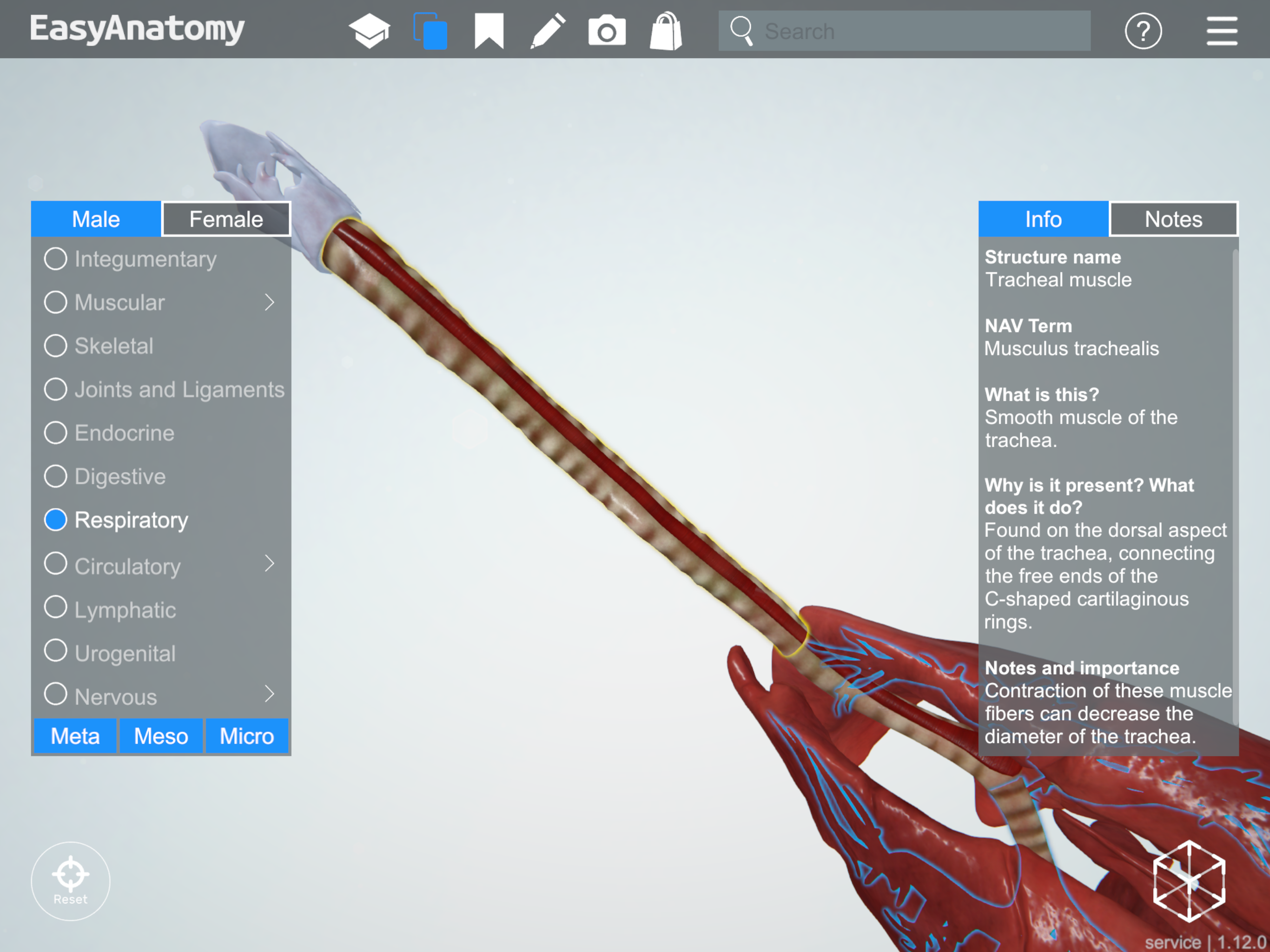
Trachea
Tracheal muscle
NAV Term: Musculus trachealis
What is this? Smooth muscle of the trachea.
Why is it present? What does it do? Found on the dorsal aspect of the trachea, connecting the free ends of the C-shaped cartilaginous rings.
Notes and importance: Contraction of these muscle fibers can decrease the diameter of the trachea.
Tracheal Muscle
Left lung
NAV Term: Pulmo sinister
What is this? The left lung has two lobes (with the cranial lobe incompletely divided into cranial and caudal portions).
Why is it present? What does it do? Each lung lobe is supplied by a lobar bronchus, and is separated from the other lobes by fissures.
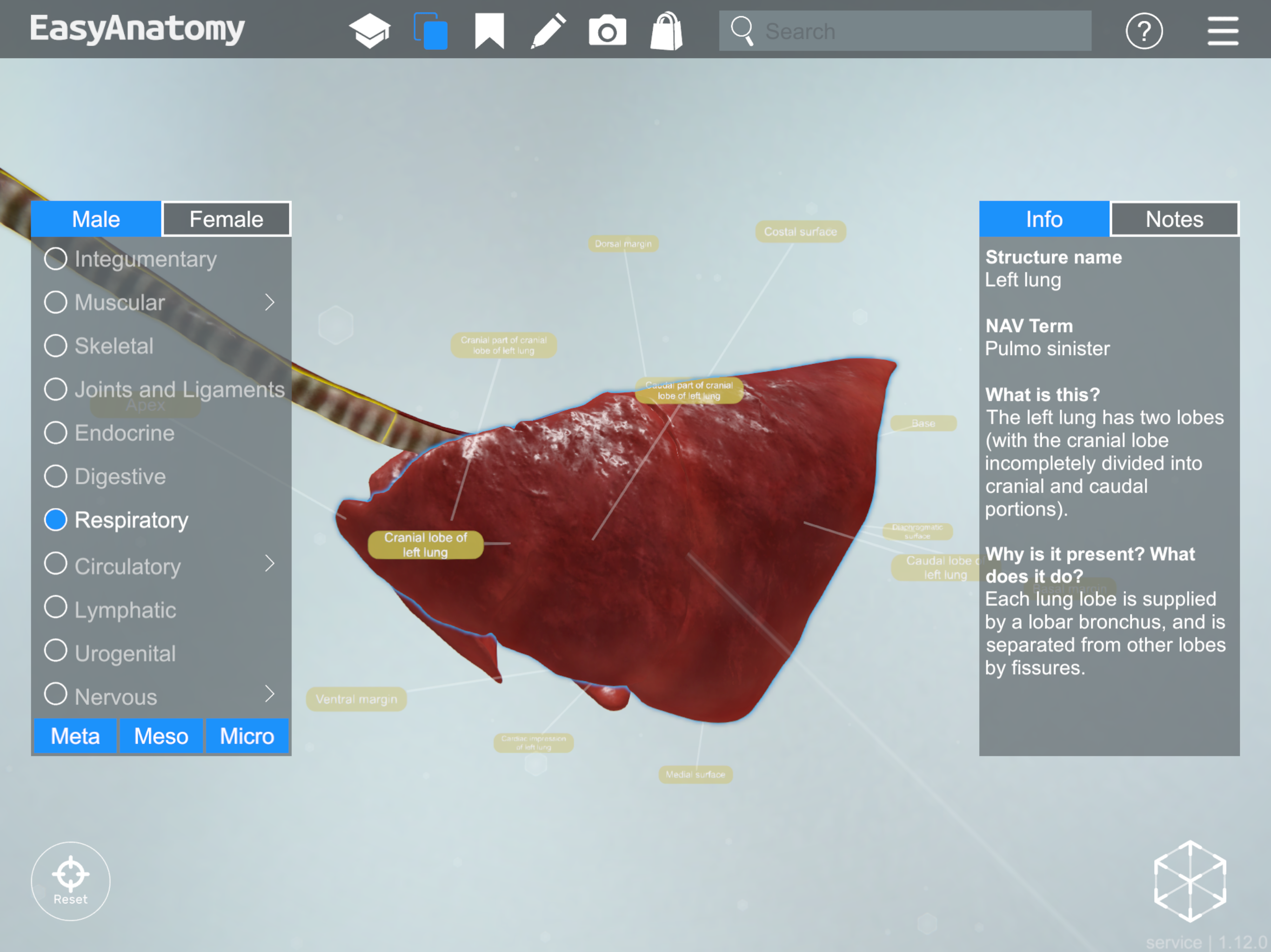
Left Lung
Right lung
NAV Term: Pulmo dexter
What is this? The right lung has four lobes: cranial, middle, caudal, and accessory.
Why is it present? What does it do? Each lung lobe is supplied by a lobar bronchus, and is separated from the other lobes by fissures.
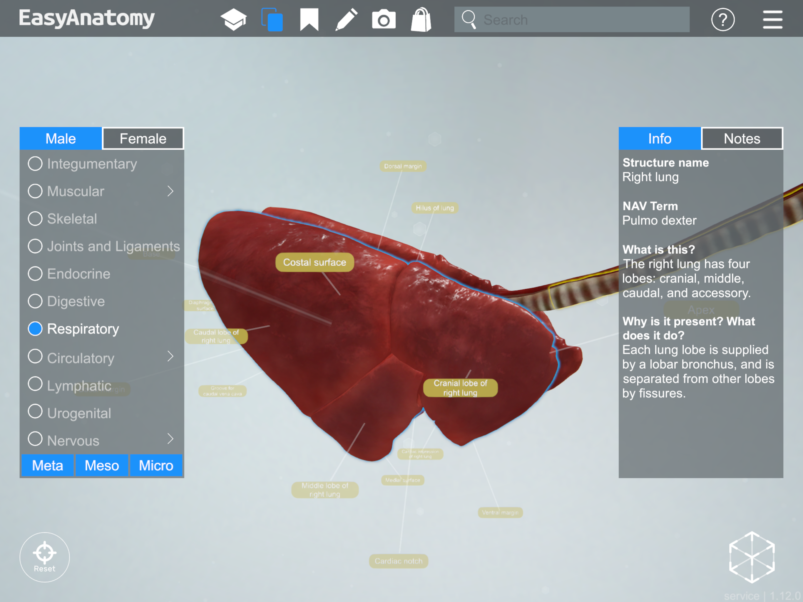
Right Lung
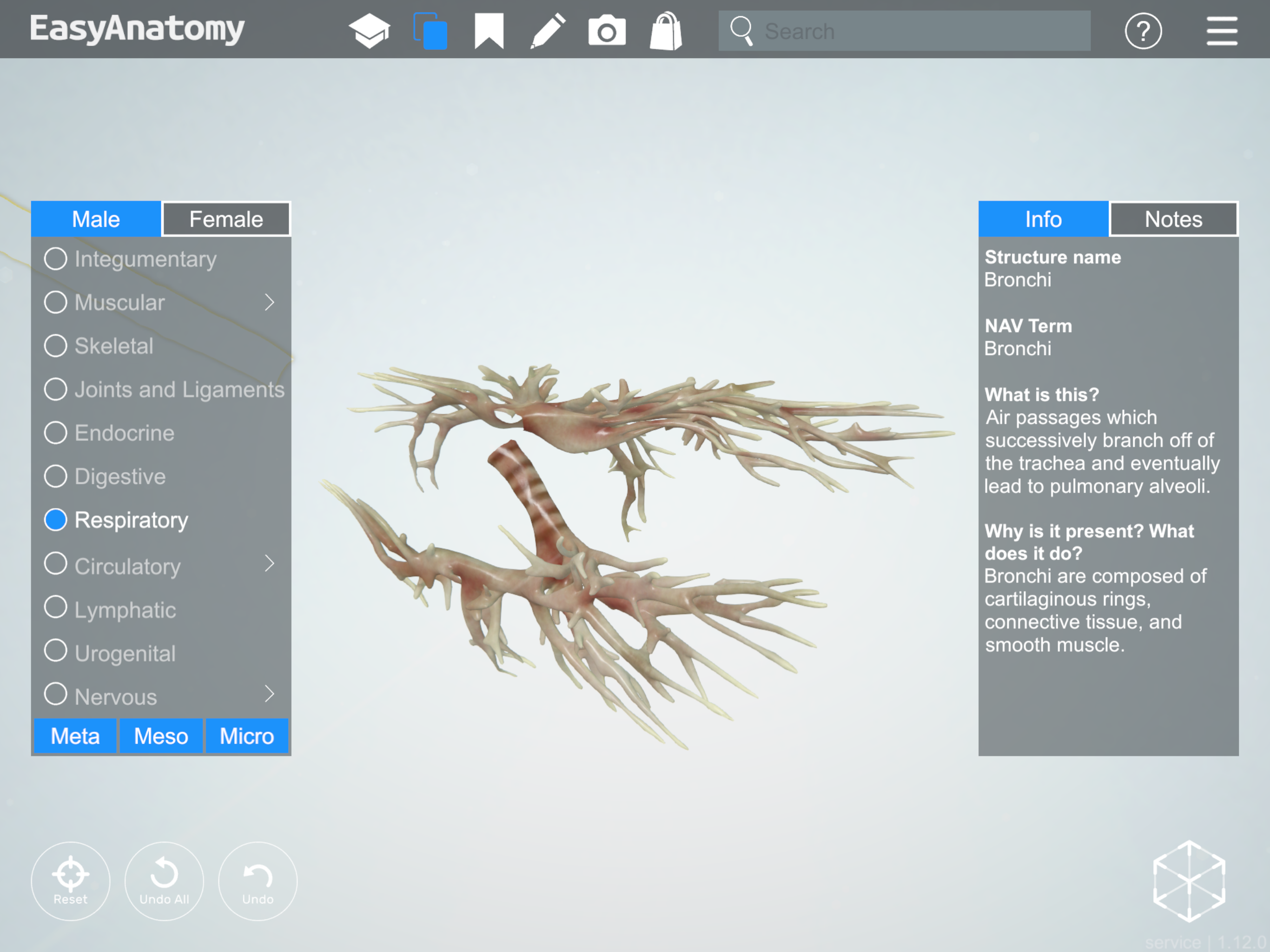
Bronchi
NAV Term: Bronchi
What is this? Air passages which successively branch off of the trachea and eventually lead to pulmonary alveoli.
Why is it present? What does it do? Bronchi are composed of cartilaginous rings, connective tissue, and smooth muscle.
Bronchi
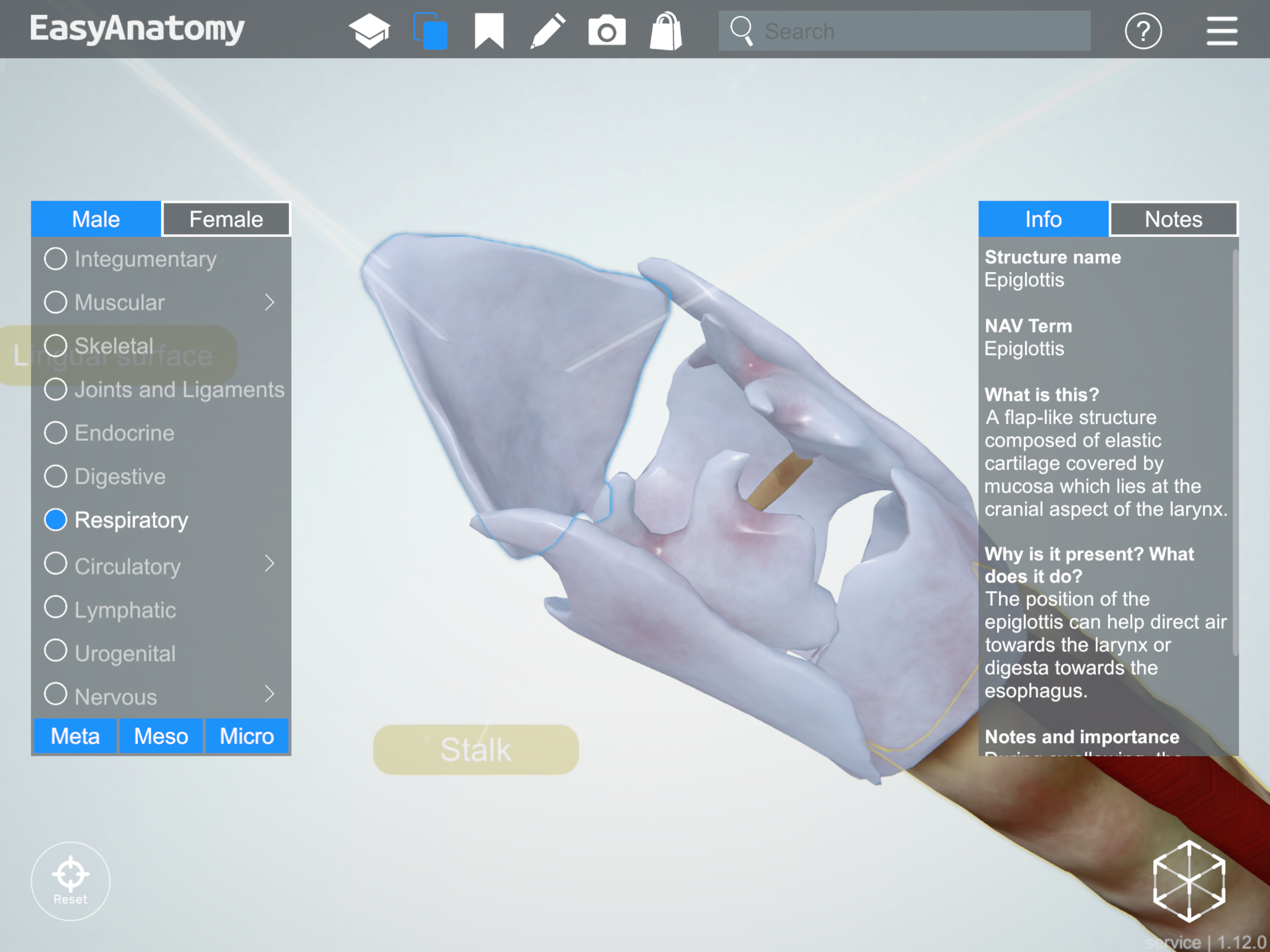
Epiglottis
NAV Term: Epiglottis
What is this? A flap-like structure composed of elastic cartilage covered by mucosa which lies at the cranial aspect of the larynx.
Why is it present? What does it do? The position of the epiglottis can help direct air towards the esophagus.
Notes and importance: During swallowing, the epiglottis moves caudally to prevent food from entering the larynx.
Epiglottis
Laryngeal Cartilages
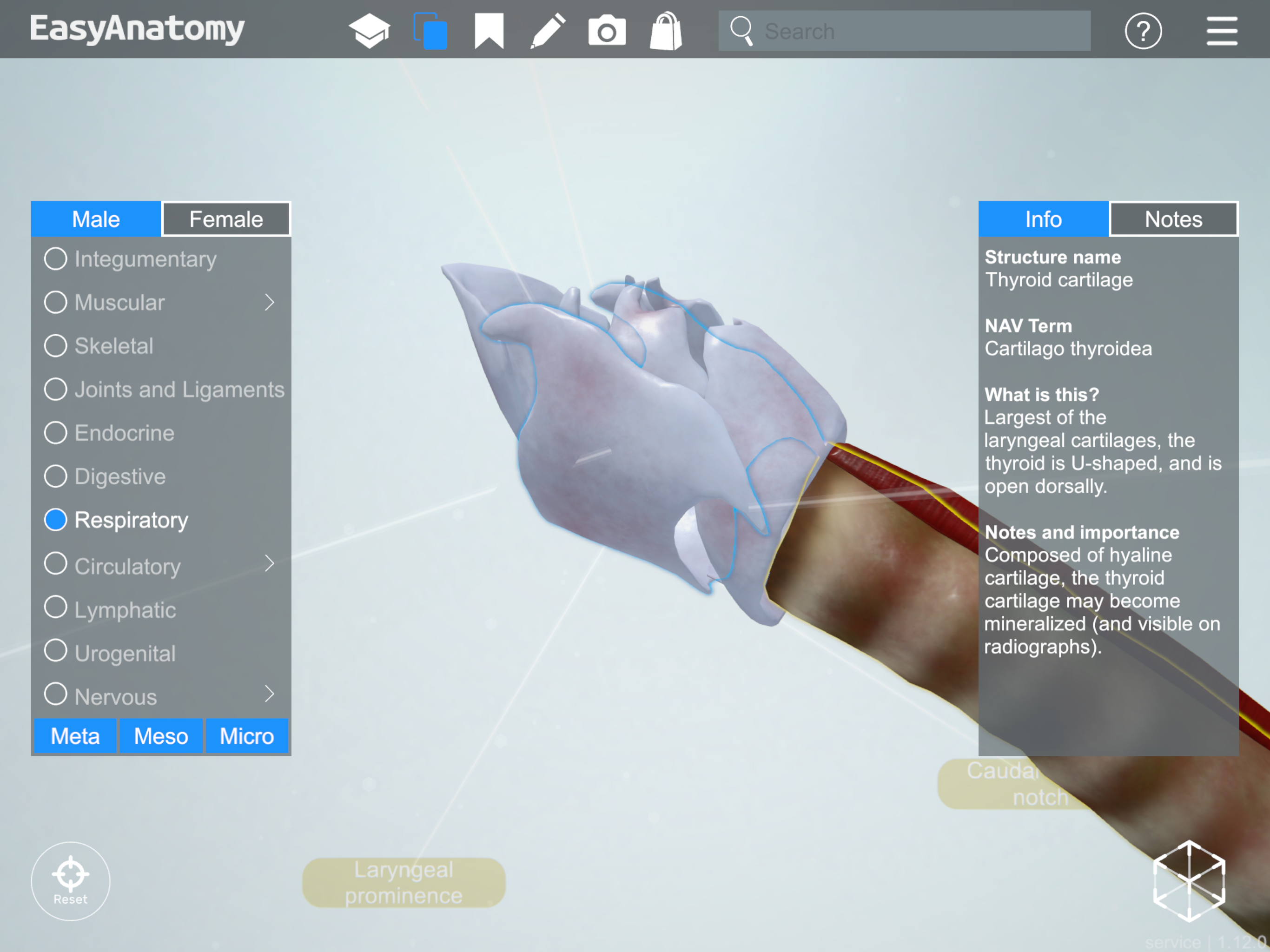
Thyroid cartilage
NAV Term: Cartilago thyroidea
What is this? Largest of the laryngeal cartilages, the thyroid is U-shaped, and is open dorsally.
Notes and importance: Composed of the hyaline cartilage, the thyroid cartilage may become mineralized (and visible on radiographs).
Thyroid cartilage
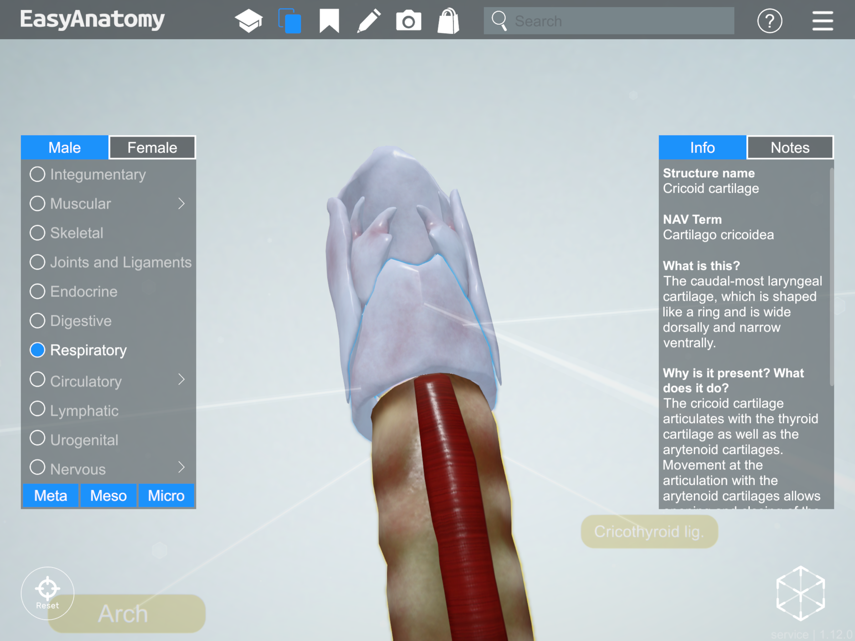
Circoid cartilage
NAV Term: Cartilago cricoidea
What is this? The caudal-most laryngeal cartilage, which is shaped like a ring and is wide dorsally and narrow ventrally.
Why is it present? What does it do? The cricoid cartilage articulates with the thyroid cartilage as well as the arytenoid cartilages. Movement at the articulation with the arytenoid cartilages allows opening and closing of the glottis. As the most caudal cartilage of the larynx, the cricoid cartilage is attached to the trachea.
Notes and importance: Composed of hyaline cartilage, the cricoid may become mineralized (and visible on radiographs).
Circoid cartilage
Arytenoid cartilage
NAV Term: Cartilago arytenoidea
What is this? Paired cartilages of the larynx, which articulate with the cricoid cartilage and attach to the vocal and vestibular folds.
Why is it present? What does it do? Movement of these cartilages allows opening and closing of the glottis.
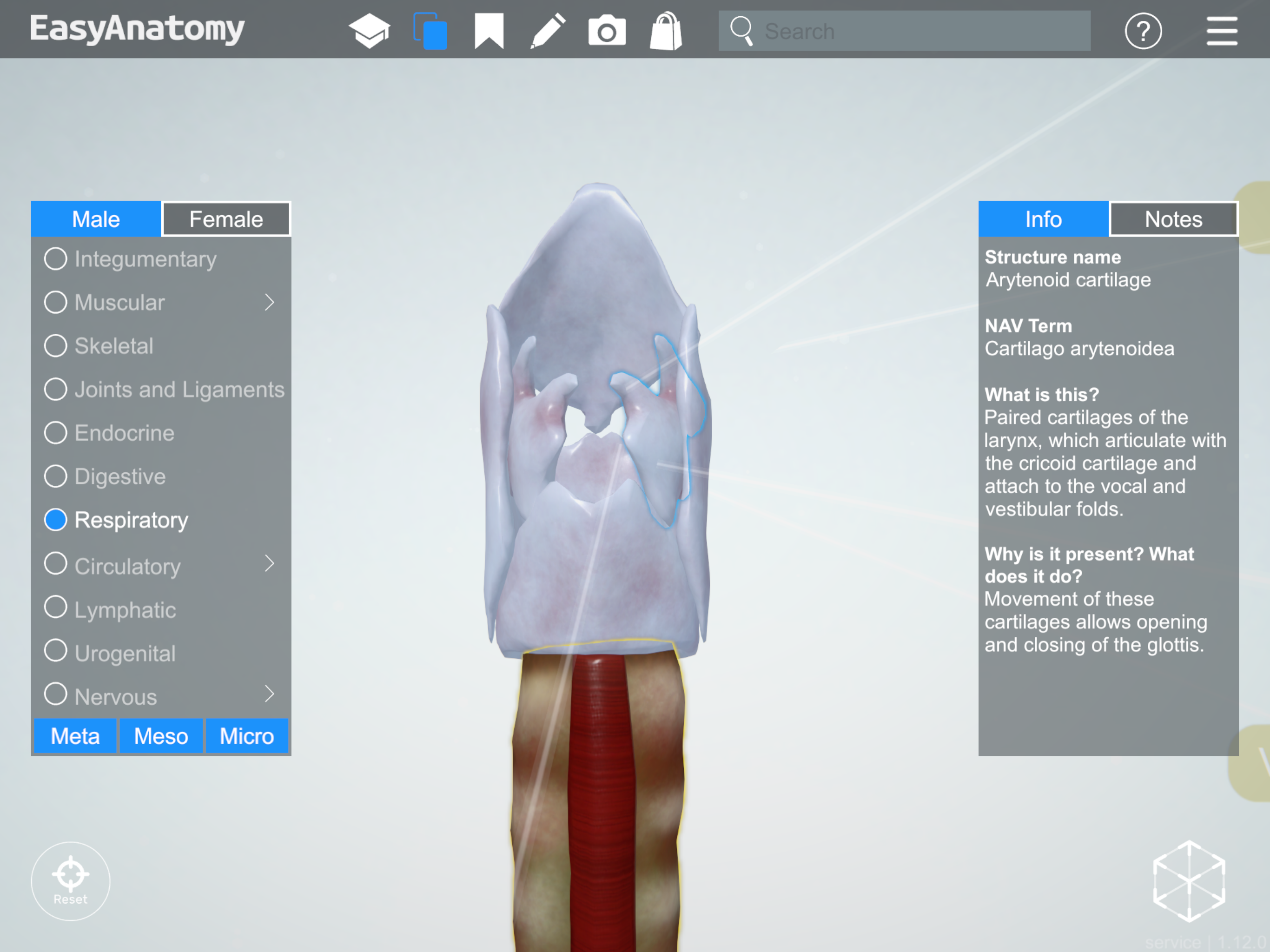
Arytenoid cartilage
—
The information and images in this post are taken from EasyAnatomy. The content is written by our team of veterinary anatomists, and our team’s goal is always to focus on what veterinary students are most likely to need to know for exams, and for clinical practice. For a similar overview on the canine knee, be sure to check out our first post in this series.
Visit our homepage to learn more about why schools such as the Western College of Veterinary Medicine, and students from over 60 schools across the world, trust EasyAnatomy for their veterinary studies – www.easy-anatomy.com.
Get Started with EasyAnatomy for FreeGet Started with EasyAnatomy for Free