Below is an overview of some of the structures that make up the male urogenital system in dogs.

Anatomy of the Canine Male Urogenital System (An Overview)
Kidney
NAV Term: Ren
What is this? Each kidney is located dorsally in the abdominal cavity (in contact with the lumbar hypaxial muscles), and is retroperitoneal in position. The right kidney is slightly more cranial (at the level of lumbar vertebrae 1-3) than the left kidney (at the level of lumbar vertebrae 2-4). The right kidney lies lateral to the caudal venal cava. The left kidney lies lateral to the aorta.
What does it do? The nephron (a specialized tubular structure) is the functional unit of the kidney, which filters blood to maintain water balance and produce urine. Kidneys also produce hormones in specialized cells (juxtaglomerular cells) of the afferent and efferent arterioles.
Notes and importance The kidneys are distinctively bean-shaped, with a rounded convex lateral border. They are surrounded by a variable amount of fat (depending on the body condition of the individual), which makes them easier to visualize on radiographs. They can be palpated (again depending on body condition), and the left kidney is easier to palpate due to its more caudal position.
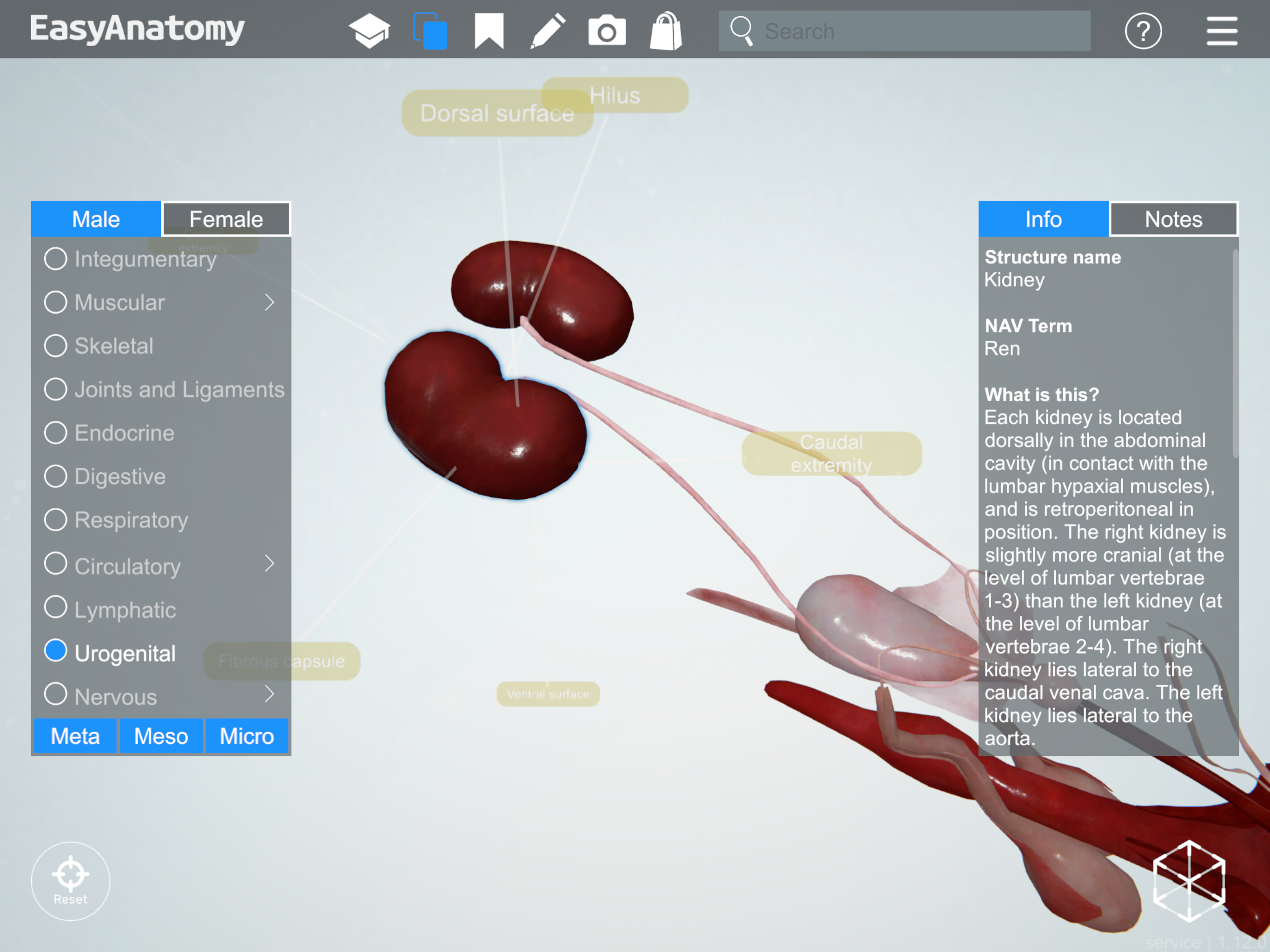
Kidney
Ureter
NAV Term: Ureter
What is this? Tubular organ which connects each kidney to the urinary bladder.
What does it do? The ureter carries urine from the kidney to the bladder. The ureters travel from dorsal to ventral as they travel from the abdominal cavity to the pelvic cavity, due to the position of the kidneys (dorsal in the abdominal cavity) and the bladder (ventral in the pelvic cavity).
Notes and importance: The ureter may develop abnormally, resulting in duplication, attaching to the vagina rather than the bladder, or even failing to form a complete patent tube. Urinary calculi can cause obstruction of the ureter.
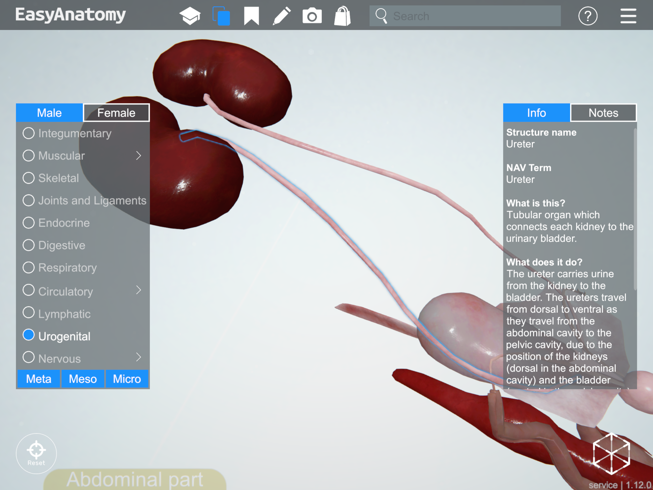
Ureter
Urinary Bladder
NAV Term: Vesica urinaria
What is this? Hollow organ located ventrally near the pelvic inlet and adjacent abdominal wall, the bladder receives urine from the ureters, and expels it via the urethra.
What does it do? Formed primarily of smooth muscle, the bladder can expand to store urine and contract to expel urine.
Notes and importance: The bladder is suspended by three folds of peritoneum, one median ligament and two lateral ligaments. The median ligament contains the remnants of the fetal urachus and umbilical arteries. The lateral ligaments contain the ureters and umbilical arteries.
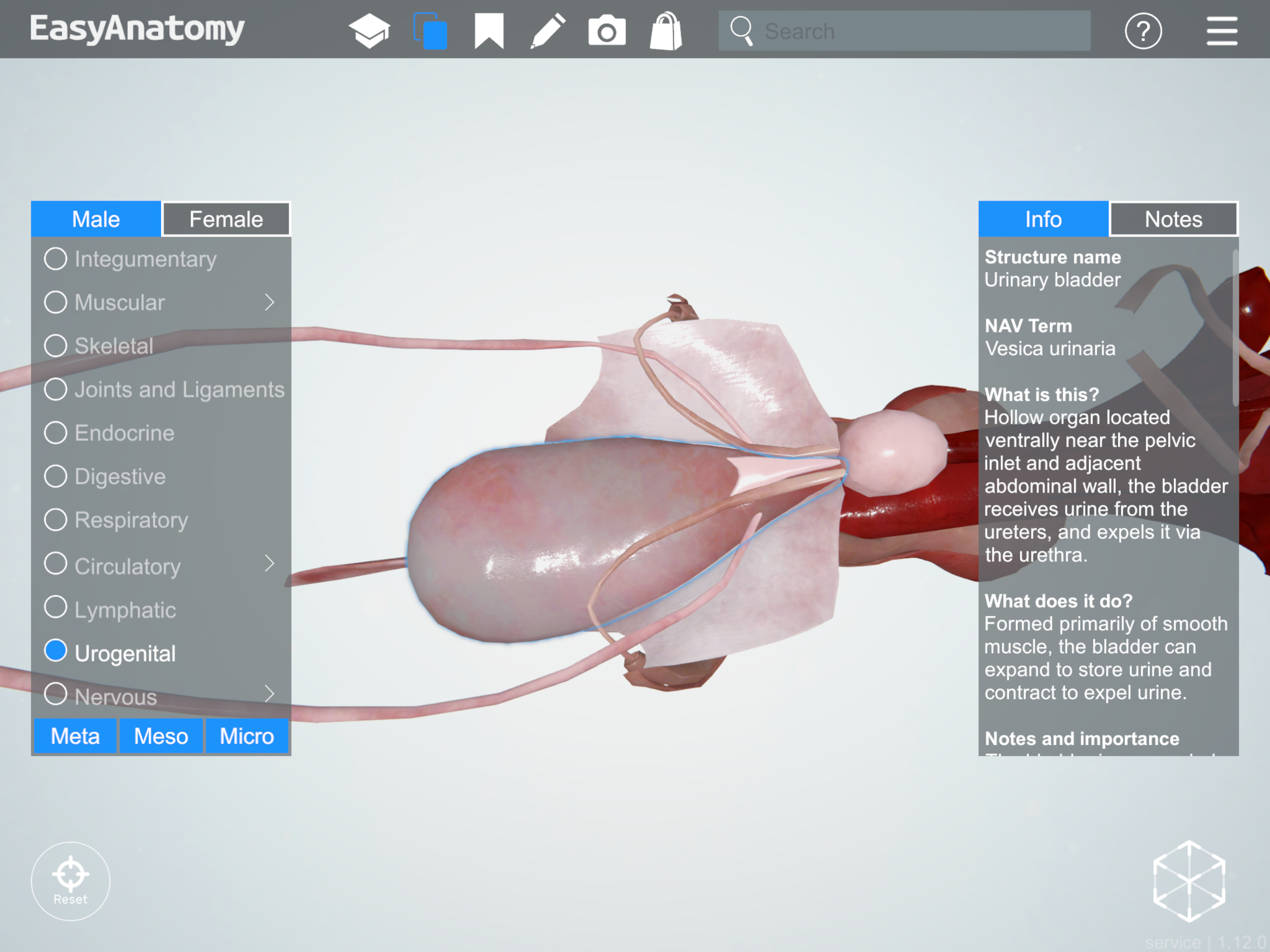
Urinary Bladder
Ductus deferens
NAV Term: Ductus deferens
What is this? Tubular organ which connects the testis/epididymis to the urethra.
What does it do? This tube carries sperm from the testis to the urethra. Because the testes are located outside the body cavity in the scrotum, the ductus deferens passes through the spermatic cord, superficial inguinal ring, the inguinal canal, and the deep inguinal ring to enter the abdominal cavity.
Notes and importance: The ductus deferens enters the urethra on its dorsal surface, after passing through the prostate gland.
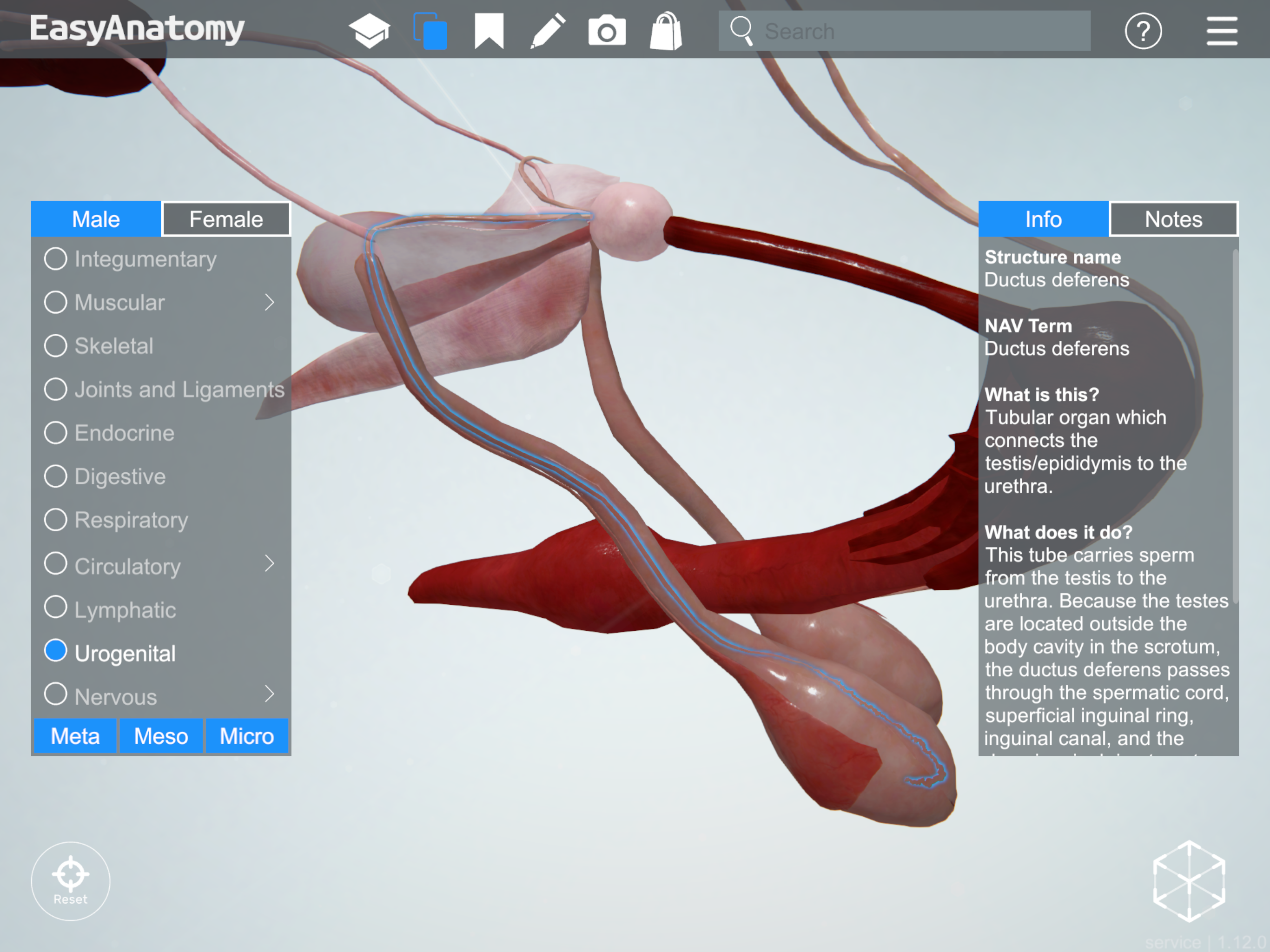
Ductus deferens
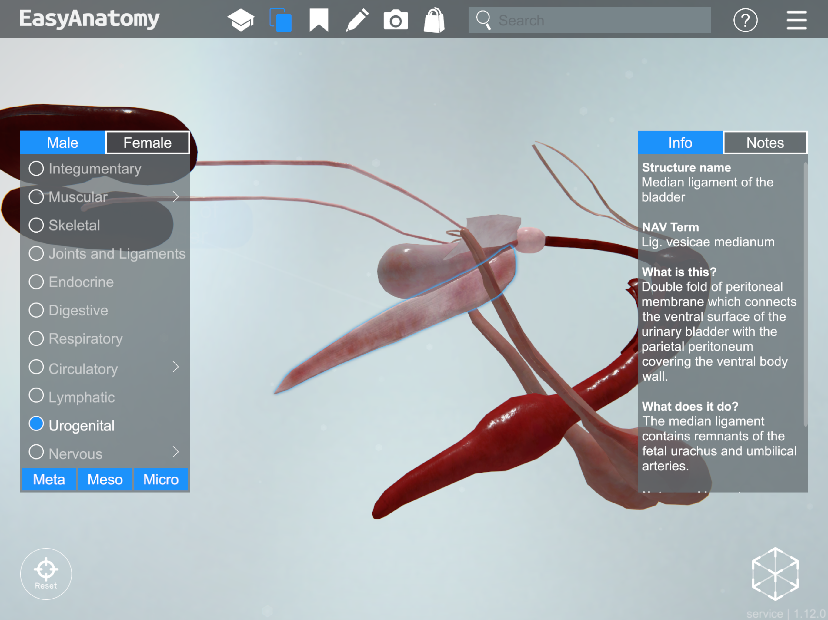
Median ligament of the bladder
NAV Term: Lig. vesicae medianum
What is this? Double fold of peritoneal membrane which connects the ventral surface of the urinary bladder with the parietal peritoneum covering the ventral body wall.
What does it do? The median ligament contains remnants of the fetal urachus and umbilical arteries.
Notes and importance: The median ligament extends cranially to the umbilicus.
Median ligament of the bladder
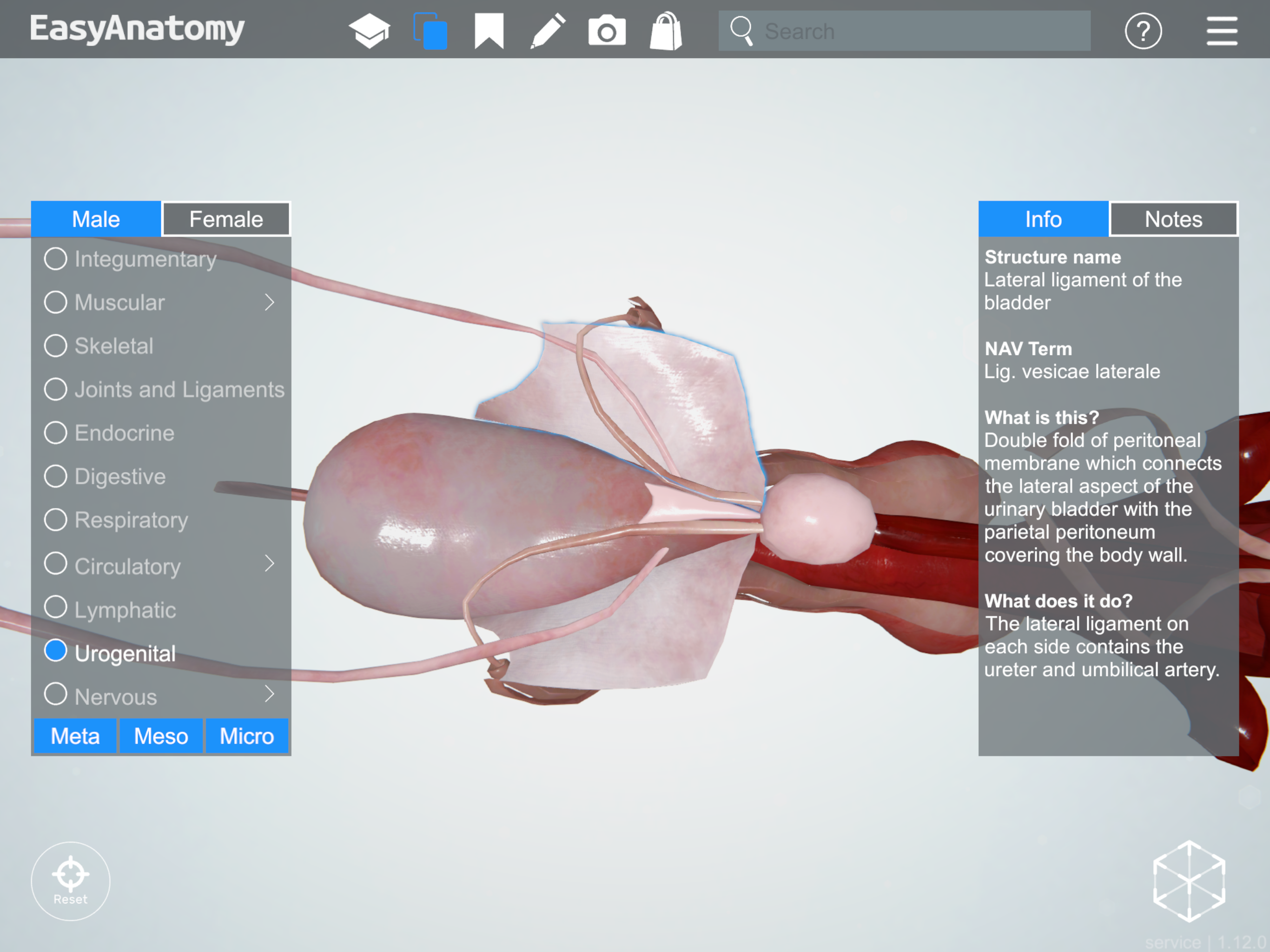
Lateral ligament of the bladder
NAV Term: Lig. vesicae laterale
What is this? Double fold of peritoneal membrane which connects the lateral aspect of the urinary bladder with the parietal peritoneum covering the body wall.
What does it do? The lateral ligament on each side contains the ureter and umbilical artery.
Lateral ligament of the bladder
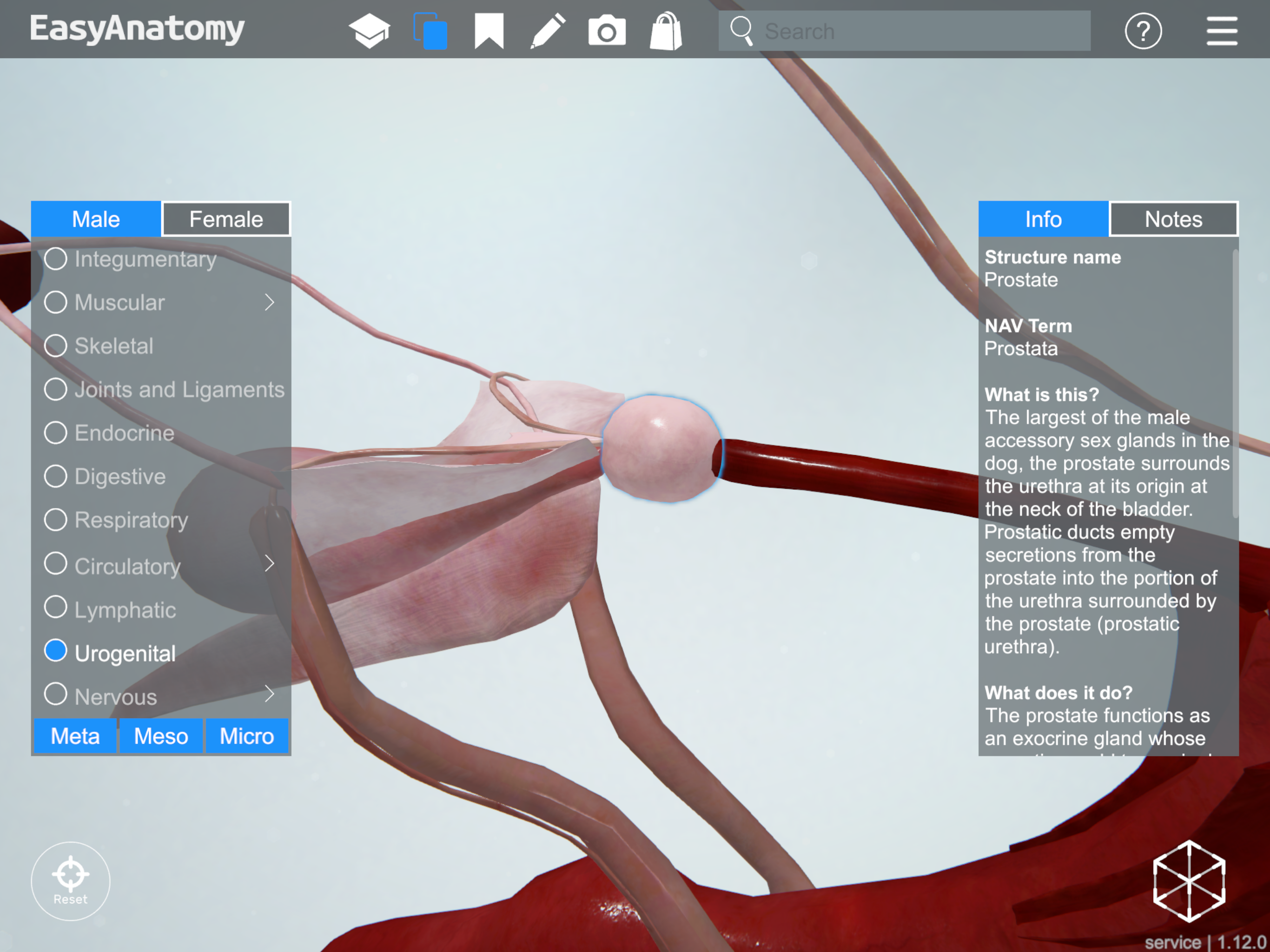
Prostate
NAV Term: Prostata
What is this? The largest of the male accessory sex glands in the dog, the prostate surrounds the urethra at its origin at the neck of the bladder. Prostatic ducts empty secretions from the prostate into the portion of the urethra surrounded by the prostate (prostatic urethra).
What does it do? The prostate functions as an exocrine gland whose secretions add to seminal fluid. Because it responds to testosterone, its size will vary greatly depending on the age and neuter status of the dog.
Notes and importance: The ductus deferens passes through the dorsal aspect of the prostate gland before entering the urethra. Enlargement of the prostate due to neoplasia or infection may occlude the urethra or the rectum, causing difficulties with urination or defecation.
Prostate
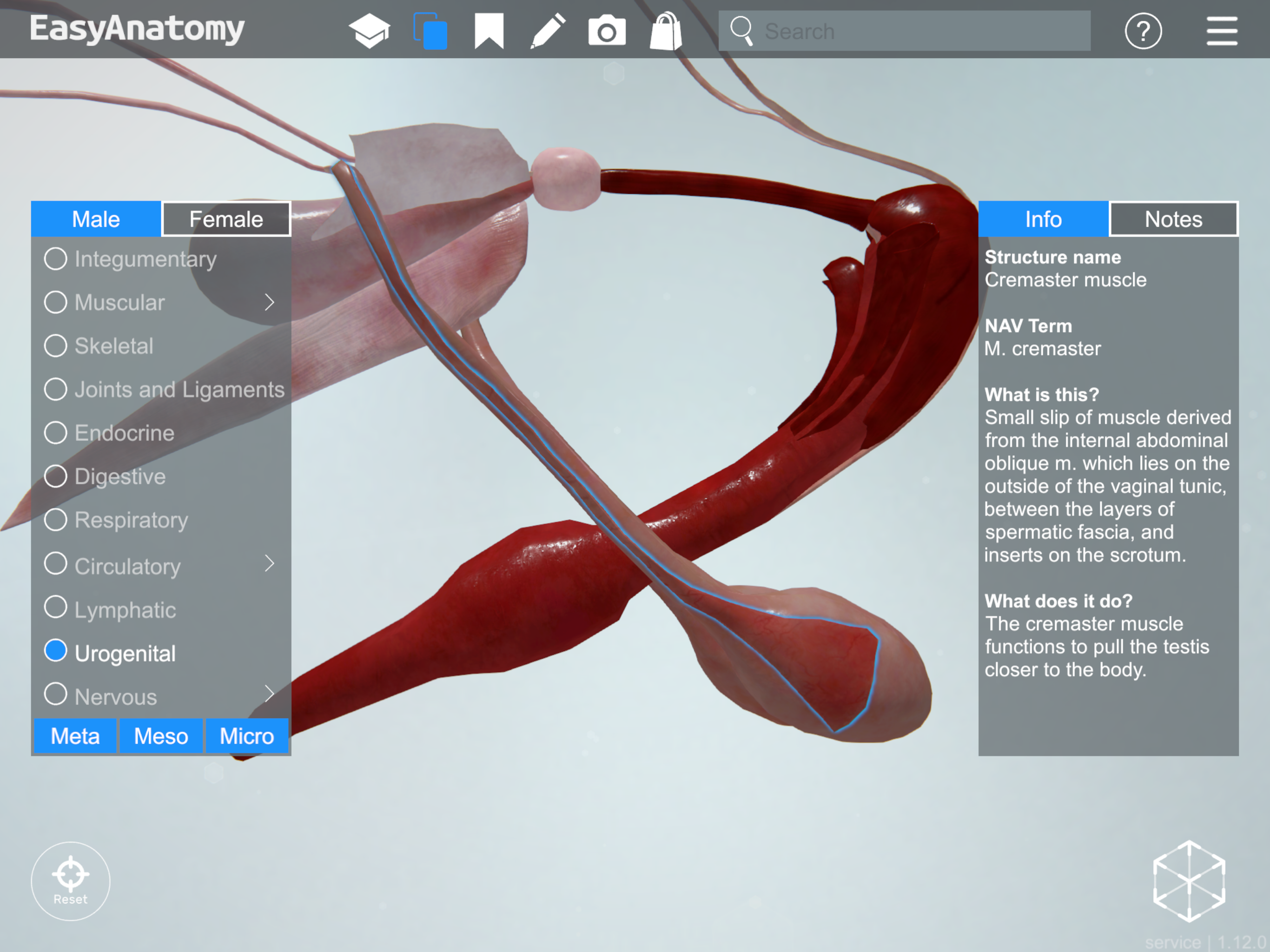
Cremaster muscle
NAV Term: M. cremaster
What is this? Small slip of muscle derived from the internal abdominal oblique m. which lies on the outside of the vaginal tunic, between the layers of spermatic fascia, and inserts on the scrotum.
What does it do? The cremaster muscle functions to pull the testis closer to the body.
Cremaster muscle
Vaginal tunic
NAV Term: Tunica vaginalis
What is this? Blind-ended outpouching of parietal peritoneum which travels out the inguinal rings and canal onto the scrotum, and wraps around the testes.
What does it do? The vaginal tunic forms during the descent of the testes, and encloses the vaginal cavity (an extension of the peritoneal cavity).
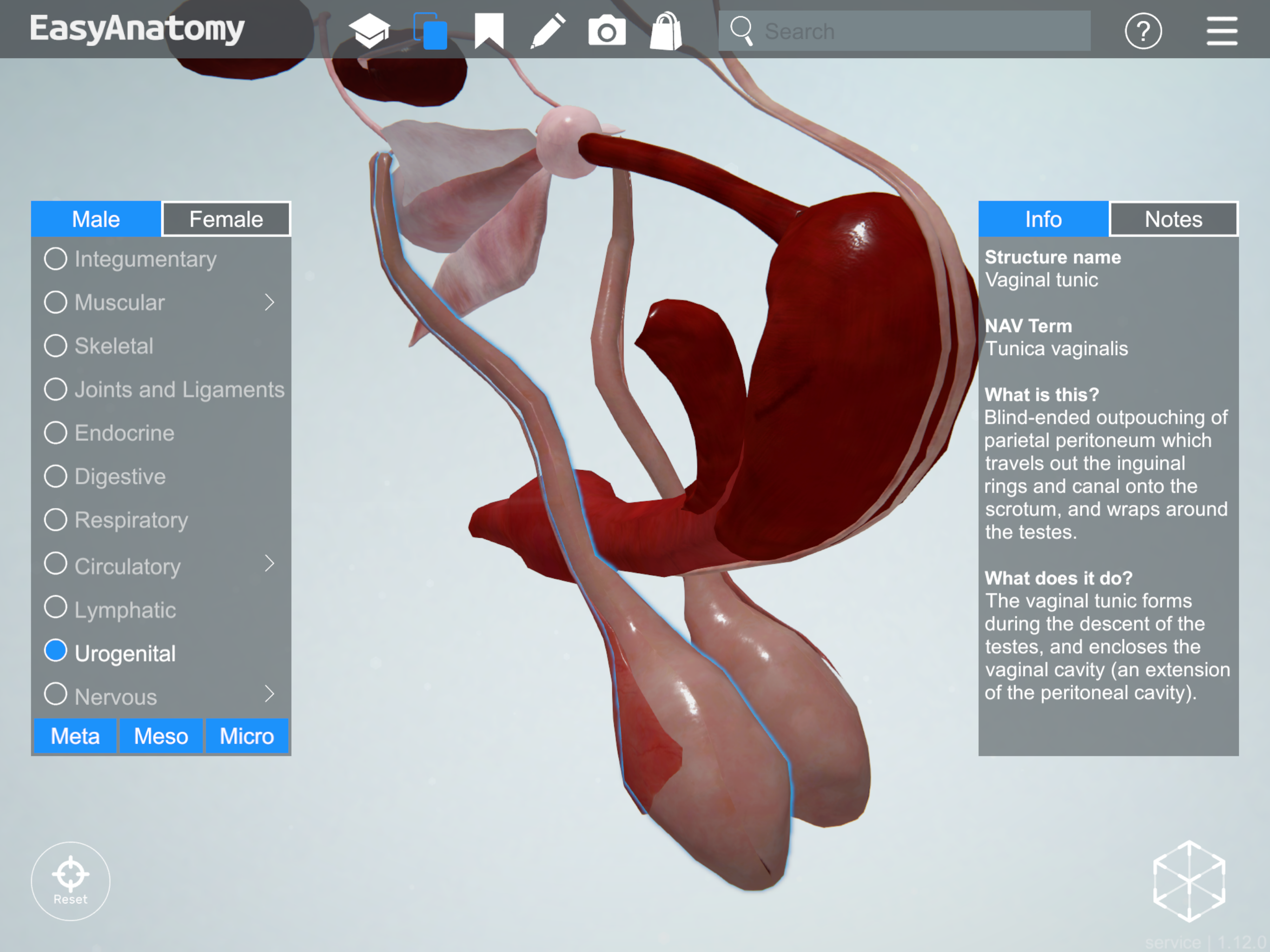
Vaginal tunic
Penis
NAV Term: Penis
What is this? The male intromittent organ, composed of vascular (erectile) tissue, smooth muscle, and striated muscle, as well as the urethra.
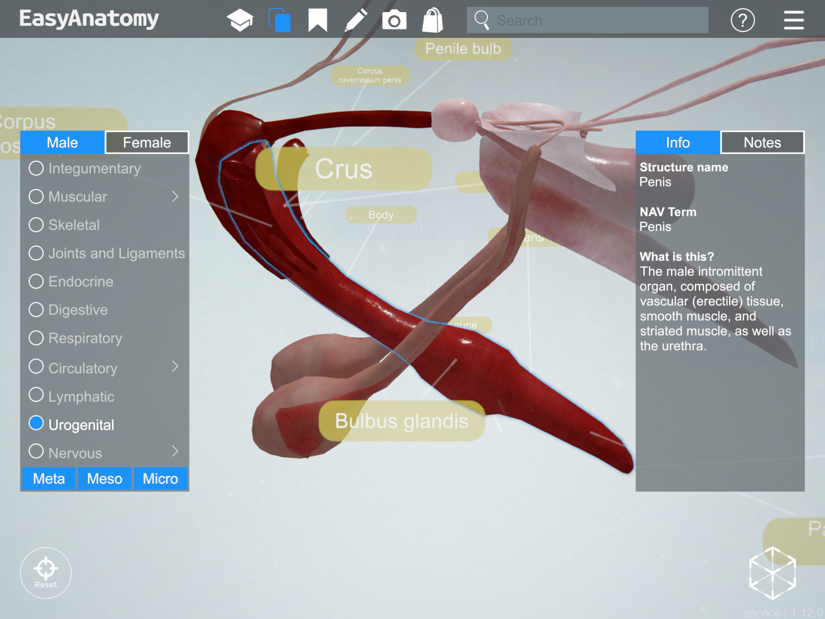
Penis
- Os Penis
- What is this? Bone found within the penis, located cranial to the corpus cavernosum. The apex extends cranially to the tip of the penis cia a cartilaginous extension.
- What does it do? The urethra (surrounded by corpus spongiosum) travels in a groove in the ventral surface of the os penis.
- Notes and importance: The os penis is attached to the distal (cranial) portion of the corpus cavernosum, and initially develops as a fibrous outgrowth from that structure.
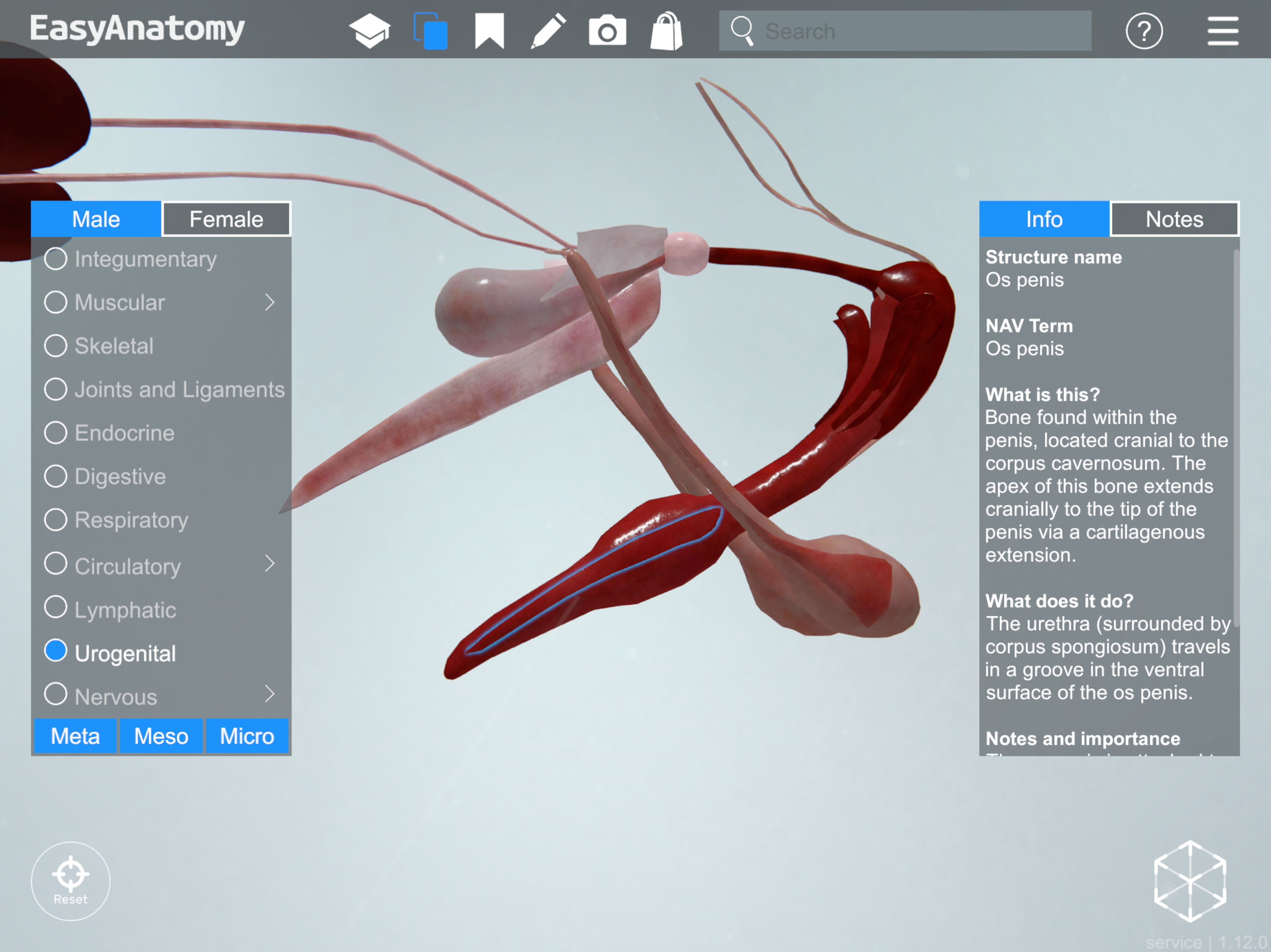
Os penis
- Penile Bulb
- What is this? Expansion of the corpus spongiosum at the proximal end of the penis, covered by bulbospongiosus muscle.
- What does it do? The penile bulb is one component of the root of the penis (along with the right and left crura)
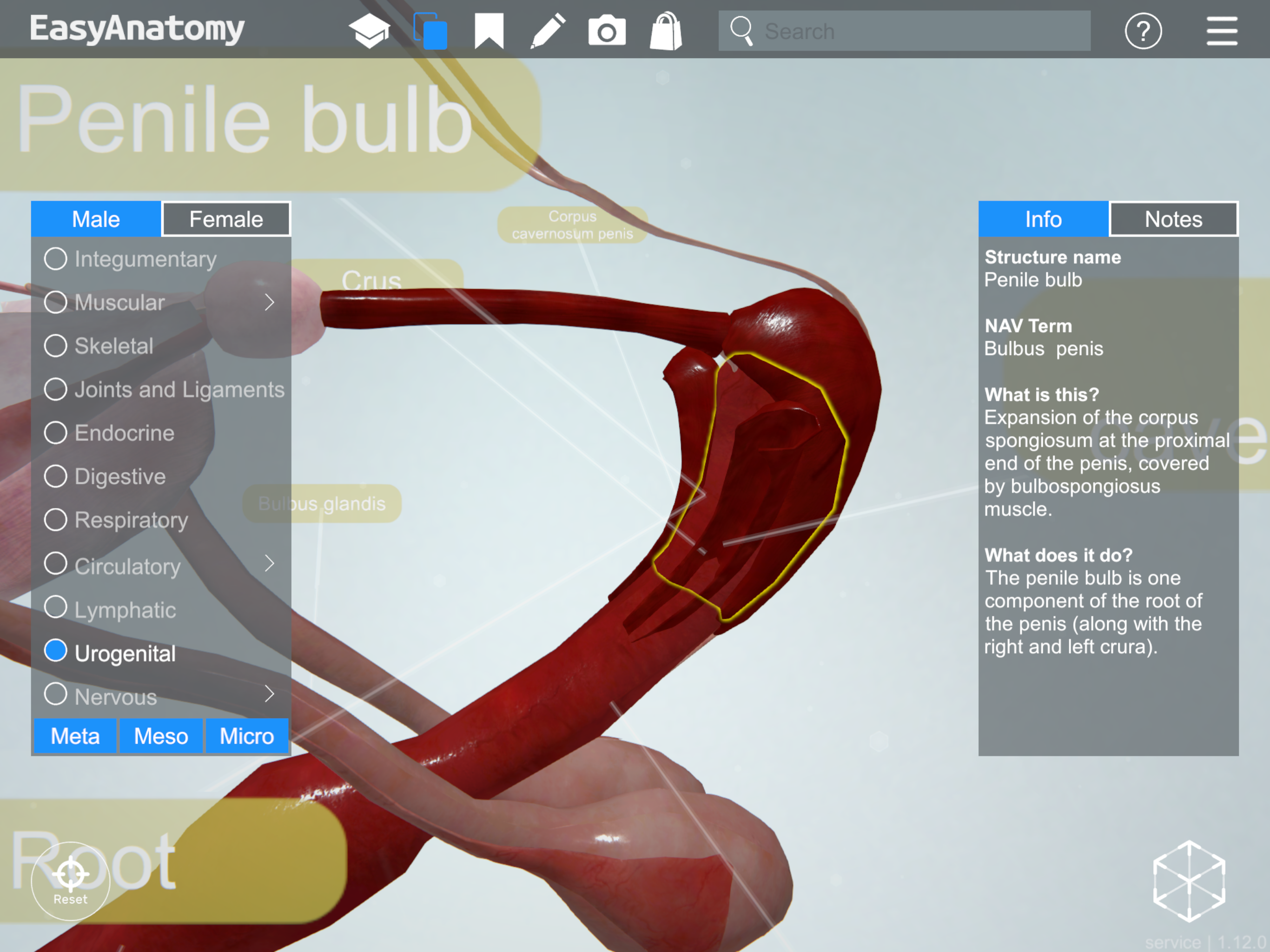
Penile bulb
- Glans
- What is this? Distal part of the penis which expands during erection.
- Notes and importance: The glans in the dog has two distinct parts, the more proximal bulbus glandis and the distal pars longa glandis
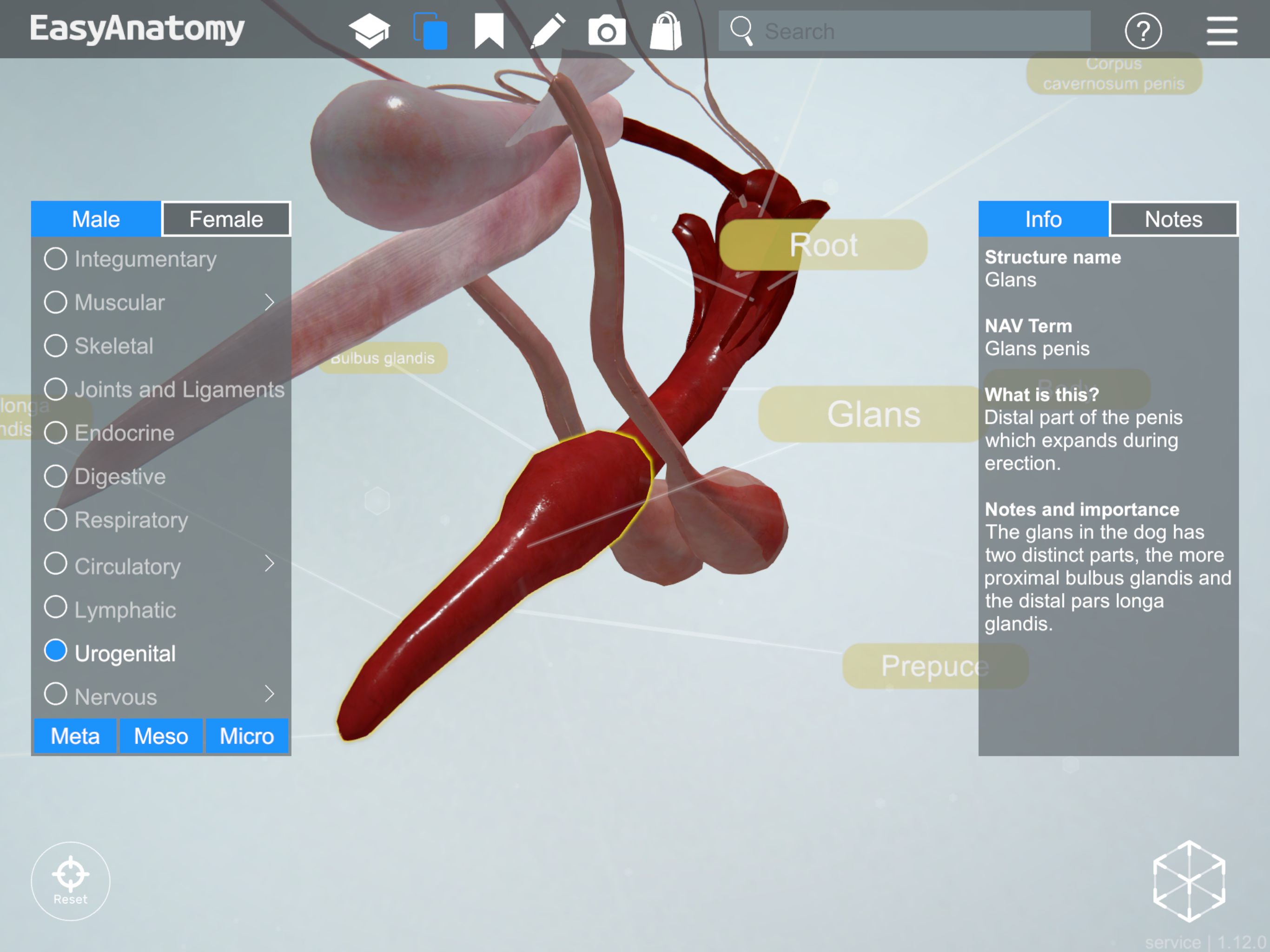
Glans
- Root
- What is this? The proximal region of the penis, composed of the two crura plus the bulb of the penis (penile bulb).
- Notes and importance: The two corpora cavernosa as well as the corpus spongiosum contribute to the root of the penis.
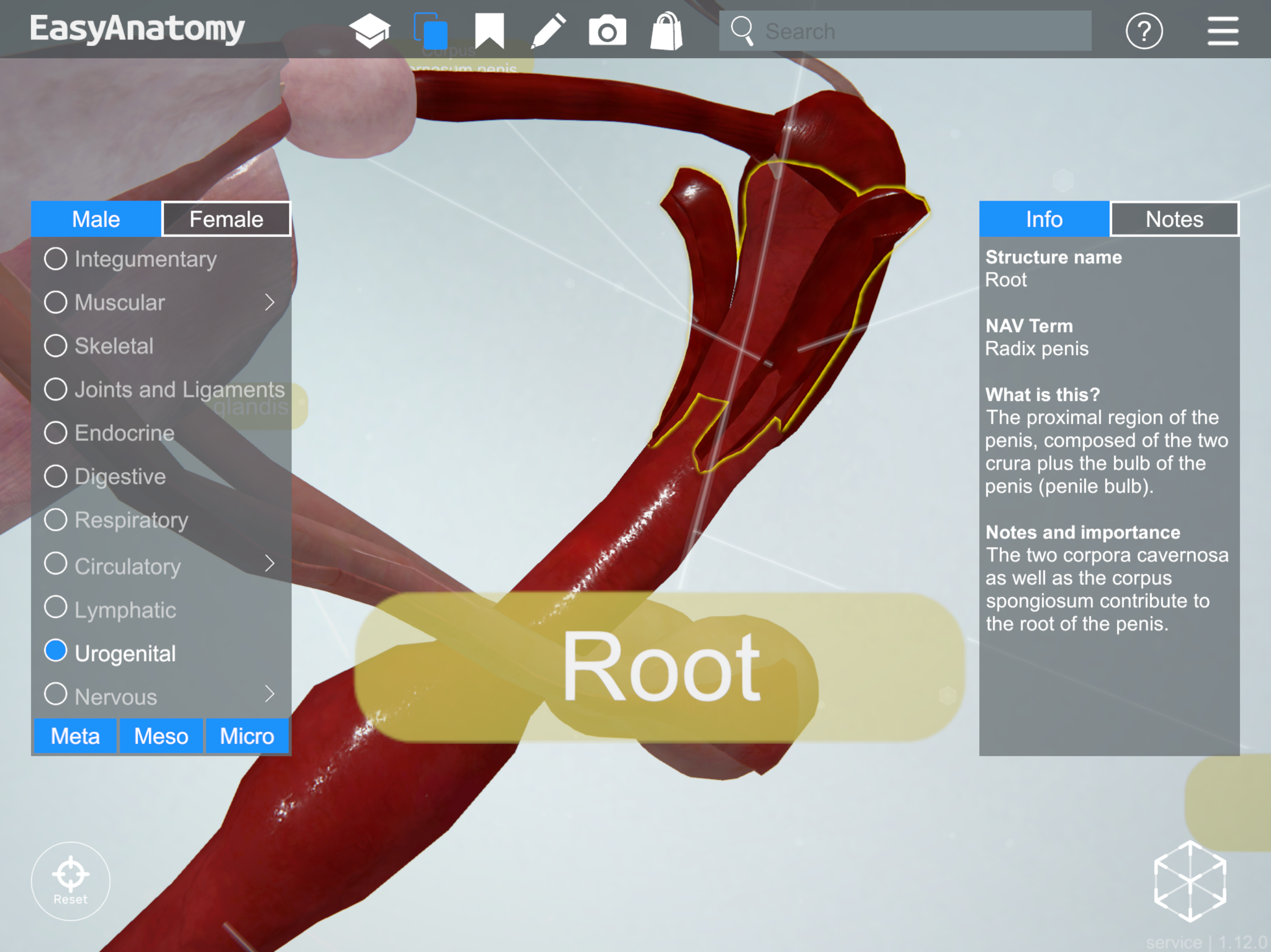
Root
- Corpus cavernosum penis
- What is this? Paired erectile tissue of the penis , which attaches proximally to the ischium and is continued distally as the os penis.
- What does it do? The corpora cavernosa contribute to the root of the penis, and form the bulk of the body of the penis. Unlike the corpus spongiosum, the corpus cavernosum is not present in the more cranial free part of the penis.
- Notes and importance: The os penis is attached to the distal (cranial) portion of the corpus cavernosum, and initially develops as a fibrous outgrowth from that structure.

Corpus cavernosum penis
Retractor penis muscle
NAV Term: M. retractor penis
What is this? Paired strap muscle which takes origin from the caudal vertebrae, loops around the anus, and travels along the ventral aspect of the penis and insert at the level of the preputial fornix.
What does it do? Contraction of this muscle maintains the position of the non-erect penis within the prepuce.
Notes and importance: The retractor penis muscle consists of smooth muscle fibers, while the other extrinsic muscles of the penis are striated muscle.
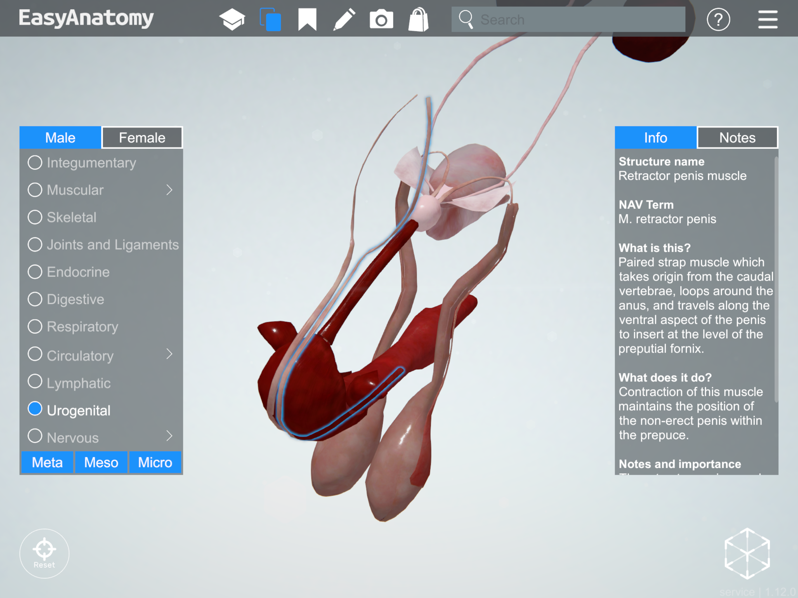
Retractor penis muscle
Ischiocavernosus muscle
NAV Term: M. ischiocavernosus
What is this? Striated muscle which covers each crus. Takes origin from the ischial tuberosity and inserts on the proximal region of the corpus cavernosum.
What does it do? Contraction of this muscle forces blood into the corpora cavernosum during erection, and prevents blood from draining from the erectile tissue.
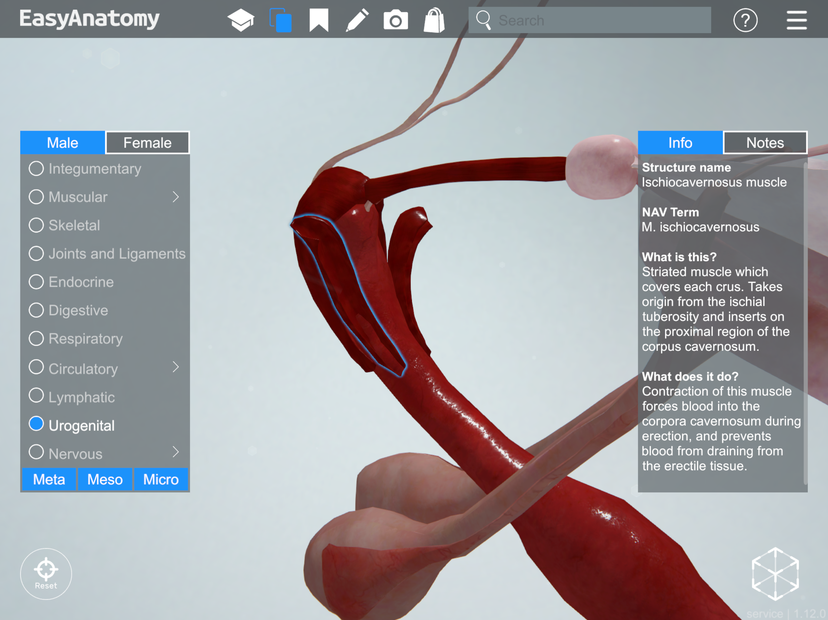
Ischiocavernosus muscle
Bulbospongiosus muscle
NAV Term: M. bulbospongiosum
What is this? Striated muscle which lies over the bulb of the penis, arising from the tunica albuginea on each side of the corpus spongiosum.
What does it do? Contraction of this muscle forces blood into the corpus spongiosum during erection, and prevents blood from draining from the erectile tissue.
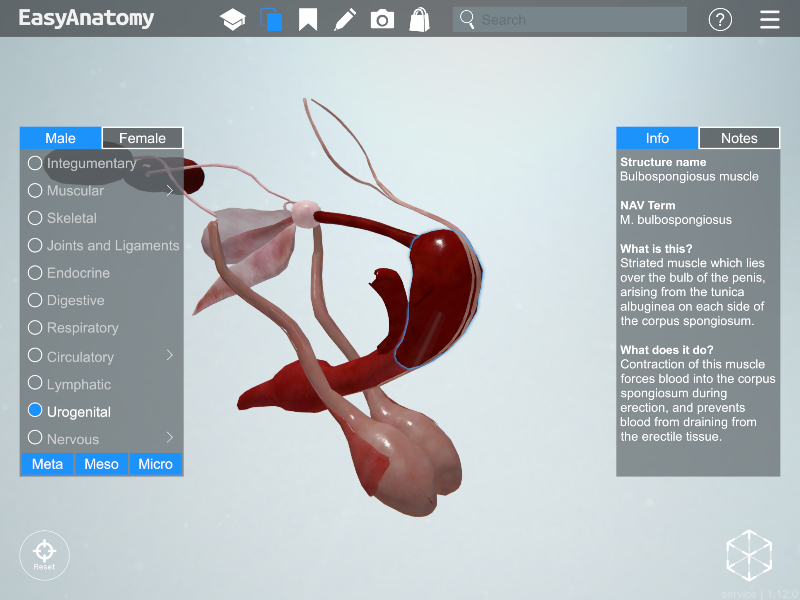
Bulbospongiosus muscle
—
The information and images in this post are taken from EasyAnatomy. The content is written by our team of veterinary anatomists, and our team’s goal is always to focus on what veterinary students are most likely to need to know for exams, and for clinical practice. For a similar overview on the canine knee, be sure to check out our first post in this series.
Visit our homepage to learn more about why schools such as the Western College of Veterinary Medicine, and students from over 60 schools across the world, trust EasyAnatomy for their veterinary studies – www.easy-anatomy.com.
Get Started with EasyAnatomy for FreeGet Started with EasyAnatomy for Free