It’s no secret that dogs love food. This love of food often leads them to eat pretty well whatever they can get their paws on, meaning stomach problems and swallowed objects are things you will commonly encounter as a veterinarian. To make sure you’re prepared when the inevitable post-snack issues arrive, a solid understanding of the anatomy of the canine digestive system is essential.

This post highlights the anatomy of the canine digestive system, and includes information such as why the structures are present, plus their importance.
The Anatomy of the Canine Digestive System (An Overview)
Esophagus
NAV Term: Esophagus
What is this? A muscular tube that carries ingesta from the laryngopharynx to the stomach. The esophagus courses through the neck, thorax, and into the abdomen. The esophagus can therefore be referred to as having cervical, thoracic, and abdominal portions.
Stomach
NAV Term: Ventriculus (Gaster)
What is this? The stomach is a dilation of the alimentary canal. It is located in the cranial region of the abdomen, caudal to the liver.
Why is it present? The stomach serves as a temporary storage site for food and produces substances that participate in digestion.
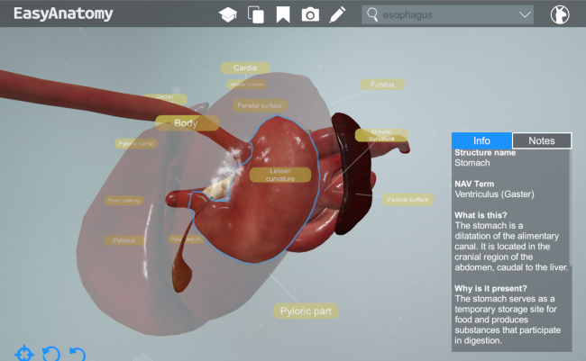
The Stomach
Notes and Importance: The dog has a simple stomach that is entirely glandular (compared tot he complex ruminant stomach that is composed of non-glandular and glandular regions). The shape and position of the stomach vary greatly depending on the amount of ingesta located within it.
Small Intestine
What is this? The initial portion of the intestinal tract, located between the stomach and large intestine. The small intestine is comprised of the duodenum, jejunum, and ileum.
Why is it present? For digestion and absorption of nutrients in the ingesta.
Notes and Importance: The average length of the small intestine is 4.0 meters (13 feet).
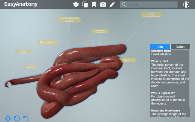
Small Intestine
- Duodenum
- What is this? The first region of the small intestine. The duodenum courses from the pylorus of the stomach to the jejunum. It is the most fixed portion of the small intestine and is located dorsally in the abdomen.
- Why is it present? For digestion and absorption of nutrients in the ingesta.
- Notes and Importance: The duodenum begins in the cranial region of the abdomen on the right side. It courses caudally on the right side, turns to the left caudal to the cranial mesenteric artery, and then courses cranially to the left of the midline.
- Jejunum
- What is this? The longest portion of the small intestine. The jejunum is the segment of intestine located between the duodenum and the ileum.
- Why is it present? For digestion and absorption of nutrients in the ingesta.
- Notes and Importance: The mesentary of the jejunum (mesojejunum) is long, allowing the jejunum to reside throughout the abdomen.
- Ileum
- What is this? The shortest portion of the small intestine. The illeum is located between the jejunum and large intestine.
- Why is it present? For digestion and absorption of nutrients in the ingesta.
- Notes and Importance: There are no grossly distinguishable features between the jejunum and ileum. In many species, the ileum is defined by a mesenteric fold that courses between the ileum and cecum, the ileocecal fold. This fold is not prominent in the dog. A common way of identifying the ileum in the dog is to identify the artery that courses along the border of the ileum that is located 180 degrees from the ileum’s attachment to its mesentary (mesoileum). This is located in the caudal region of the abdomen.
Large Intestine
What is this? The terminal portion of the intestinal tract. It courses between the small intestine (ileum) and the anus. The large intestine is comprised of the cecum, colon, rectum, and anal canal.
Why is it present? For absorption of water and nutrients.
Notes and Importance: The large intestine is shorten than the small intestine. Its average length in the dog is 0.6 meters, or 2 feet.
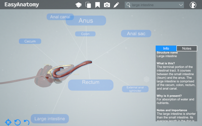
The Large Intestine
- Cecum
- What is this? A diverticulum of the initial portion of the colon (ascending colon). The cecum communicates with the ascending colon via the cecocolic orifice.
- Why is it present? The function of the cecum is the same as that of the other parts of the large intestine, primarily absorption of water.
- Notes and Importance: The cecum is located in the caudal region of the abdomen on the right side.
- Colon
- What is this? The portion of the intestinal tract located between the small intestine (ileum) and the rectum. The regions of the colon include the ascending colon, transverse colon, and descending colon.
- Why is it present? For absoprtion of water and nutrients.
- Notes and Importance: The colon is located dorsally in the abdomen. It starts on the right side and courses cranially, it then passes to the left side cranial to the root of the mesentary (around the cranial mesenteric artery), and then courses caudally along the left side of the abdomen to the pelvic cavity, where it is continued by the rectum.
- Anal Canal
- What is this? The portion of the alimentary canal located between the rectum and the anus. This region is short, approximately 1.0cm in length. It is typically located at approximately the fourth caudal vertebra.
- Notes and Importance: The anal canal is surrounded by the internal anal sphincter (smooth muscle) and the external anal sphincter (skeletal muscle). These muscles help control defecation.
Liver
NAV Term: Hepar
What is this? The liver is a large glandular organ located in the cranial part of the abdomen, between the diaphragm and stomach. The liver is comprised of the following lobes: caudate, right lateral, right medial, quadrate, left medial, and left lateral.
Why is it present? The liver produces bile (exocrine function) and substances released into the bloodstream that act on the metabolism of carbohydrates, proteins, and fats (endocrine function).
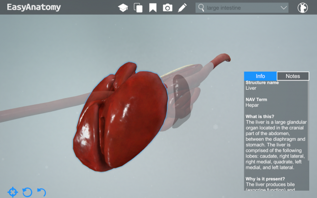
The Liver
Gall Bladder
NAV Term: Vesica Fellea
What is this? A sac-like structure located in the abdomen along the visceral surface of the liver between the quadrate and right medial lobes.
Why is it present? The gall bladder stores and concentrates bile. Bile, produced in the liver, courses to the gall bladder through hepatic ducts and then the cystic duct. When the gall bladder is stimulated to release bile, the bile travels through the cystic duct to the bile duct and into the initial portion of the duodenum.
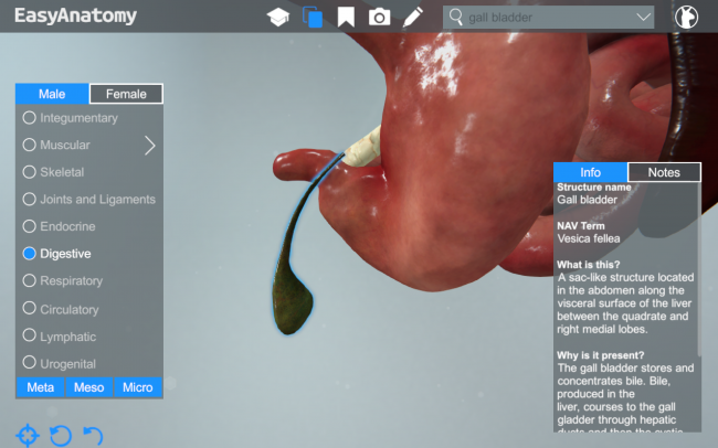
Gall Bladder
—
The information and images in this post are taken from EasyAnatomy. The content is written by our team of veterinary anatomists, and our team’s goal is always to focus on what veterinary students are most likely to need to know for exams, and for clinical practice. For a similar overview on the canine knee, be sure to check out our first post in this series.
Visit our homepage to learn more about why schools such as the Western College of Veterinary Medicine, and students from over 60 schools across the world, trust EasyAnatomy for their veterinary studies – www.easy-anatomy.com.
Get Started with EasyAnatomy for FreeGet Started with EasyAnatomy for Free